source("R/init.R")
vigns <- fs::dir_ls(
  here::here(training_dir, "Plankton_gelatinous"),
type = "file", glob = "*.png", recurse = TRUE)7 Plankton_gelatinous
7.1 Tunicata
Tunicates are a subphylum of the Chordates. Their name comes from the tunic produced by the epidermis covering them. The larval stage of these organisms has a notochord (not present in the adult stage) (Ruppert, Fox, and Barnes 2004).
7.1.1 Thaliacea
7.1.1.1 Salpida
7.1.1.1.1 salpidae
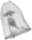
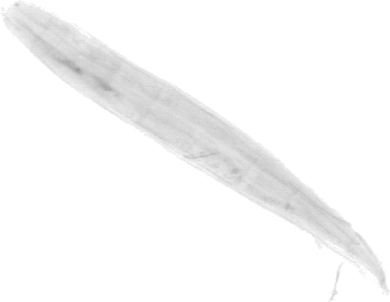
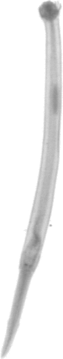
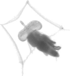
These organisms are transparent, which is indicated by the very light yellow on the vignettes. The band of muscle present in the Doliolida is not present. There is, however, the presence of muscle. The barrel shape of the Doliolida is not present. ( Section 7.1.1.3 ).
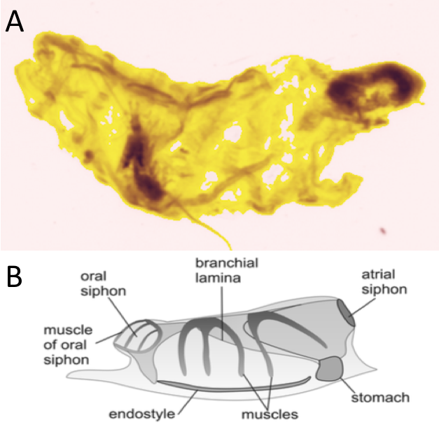
Figure 7.1 shows several vignettes of Salpidae.
plot_vignettes(vigns, group = "Salpidae")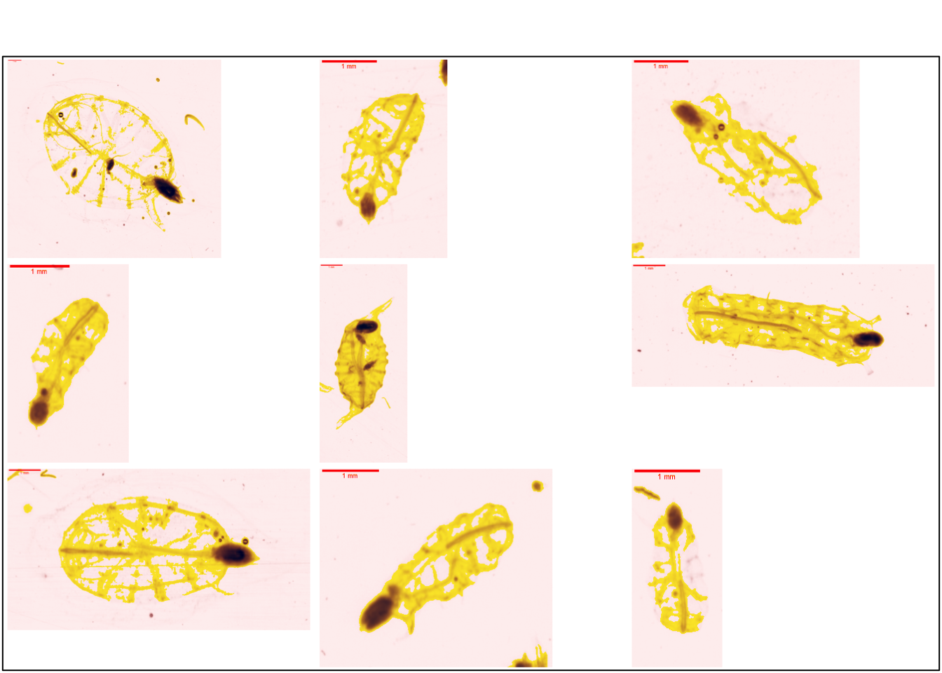
This group is similar to the salpida group in ZooScanNet (Figure 7.2).
7.1.1.2 Pyrosomatida
7.1.1.2.1 Pyrosomatida
New group
In previous studies (and associated training sets), this group was not studied.
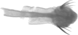
Pyrosomatida forms large tubular colonies several metres long. They are made up of thousands of blastozoids whose tunics are grouped together and the buccal siphon opens in the centre of the tube (Caicci et al. 2013).

In the ZooScanNet, they are very often found in the form of colonies (Figure 7.4). The pharynx is discernible in each image
7.1.1.3 Doliolida
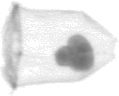
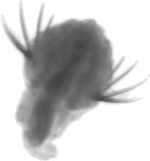
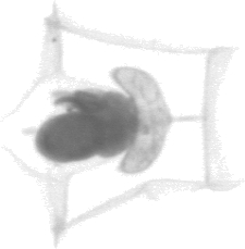
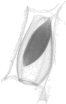
These organisms have the appearance of a symmetrical barrel with a ring of several circles (Figure 7.5). Circles are muscles surrounding the body of the organism
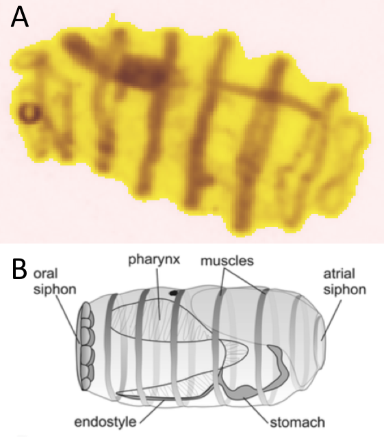
Figure 7.6 shows several vignettes of Sagittoidea. These organisms are transparent as indicated by the light yellow parts on the vignettes. The muscle bands around the organisms appear darker.
plot_vignettes(vigns, group = "Doliolida", nx = 3,ny = 2)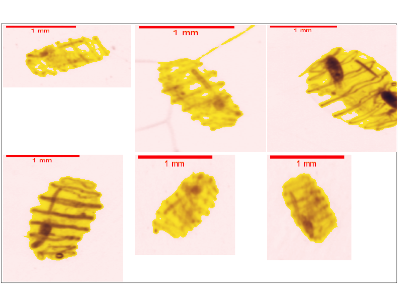
This group is similar to doliolida in ZooScanNet ( Figure 7.7 ).

7.1.2 Appendicularia
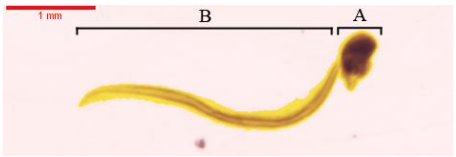
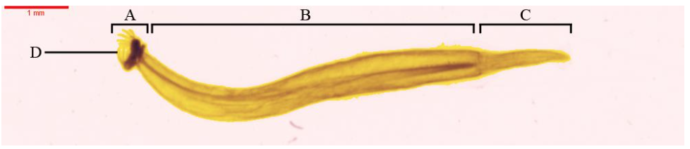
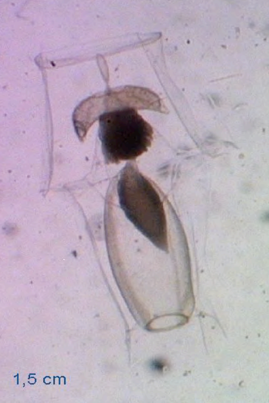
Appendidicularia keep their tails in the adult state. This neotenic character makes them easily identifiable. These organisms are divided into two parts: the trunk and the tail ( Figure 7.8 ).
7.1.2.1 Copelata
7.1.2.1.1 Oikopleuridae
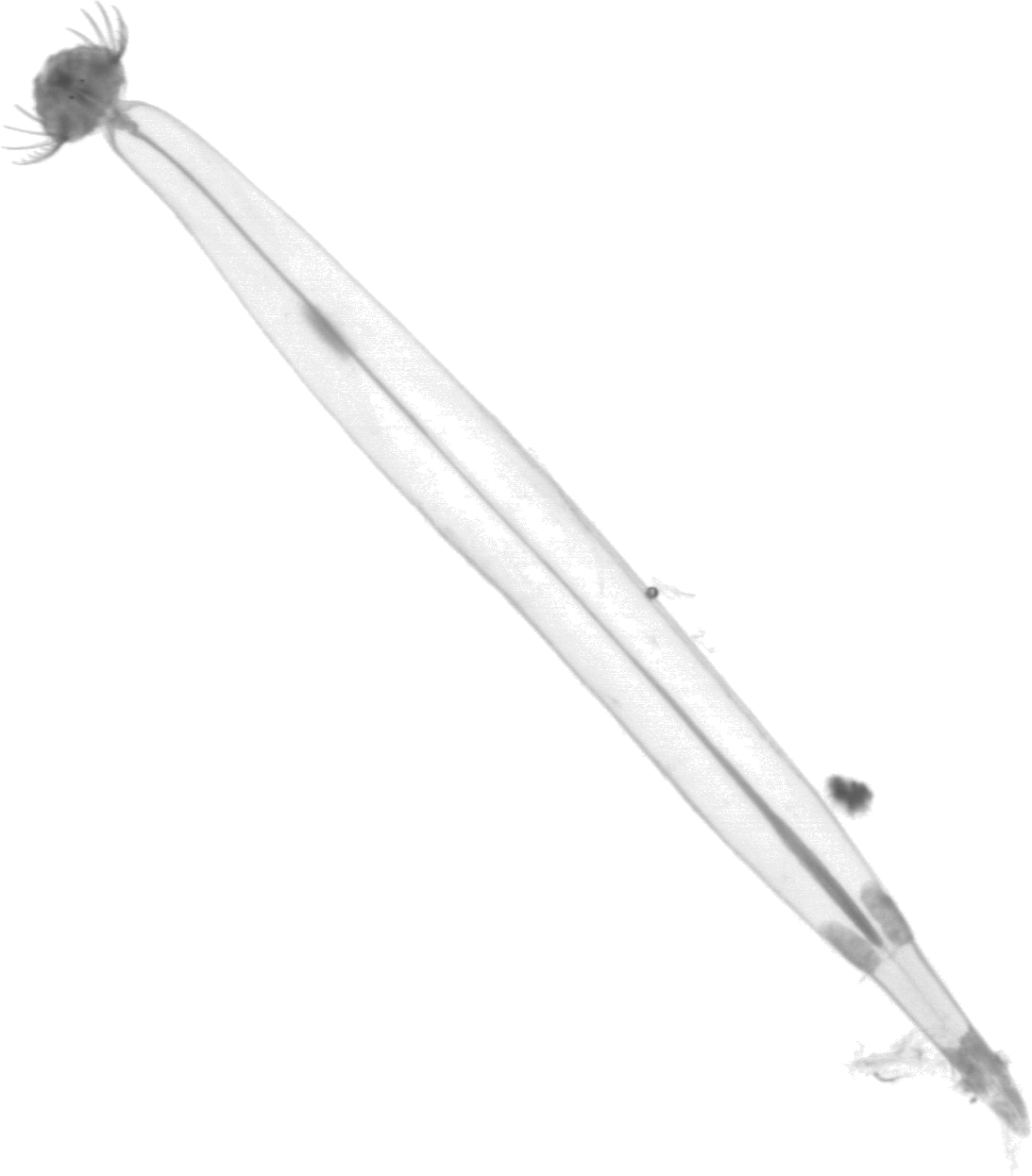
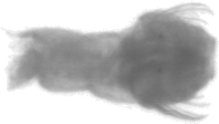
The trunk is globular and all organs are tightened. The colour of the trunk is very dark. The subchordal cells arround the notochord give a dark of the tail (Figure 7.9).
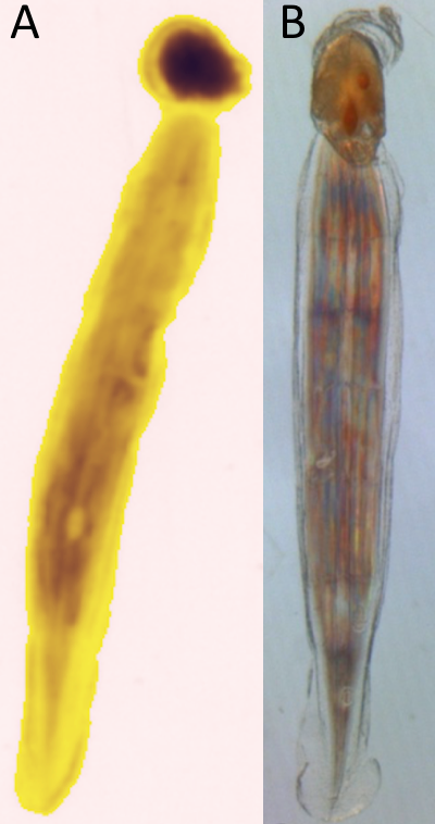
Figure 7.10 shows severals vignettes of Oikopleuridae.
plot_vignettes(vigns, group = "Oikopleuridae")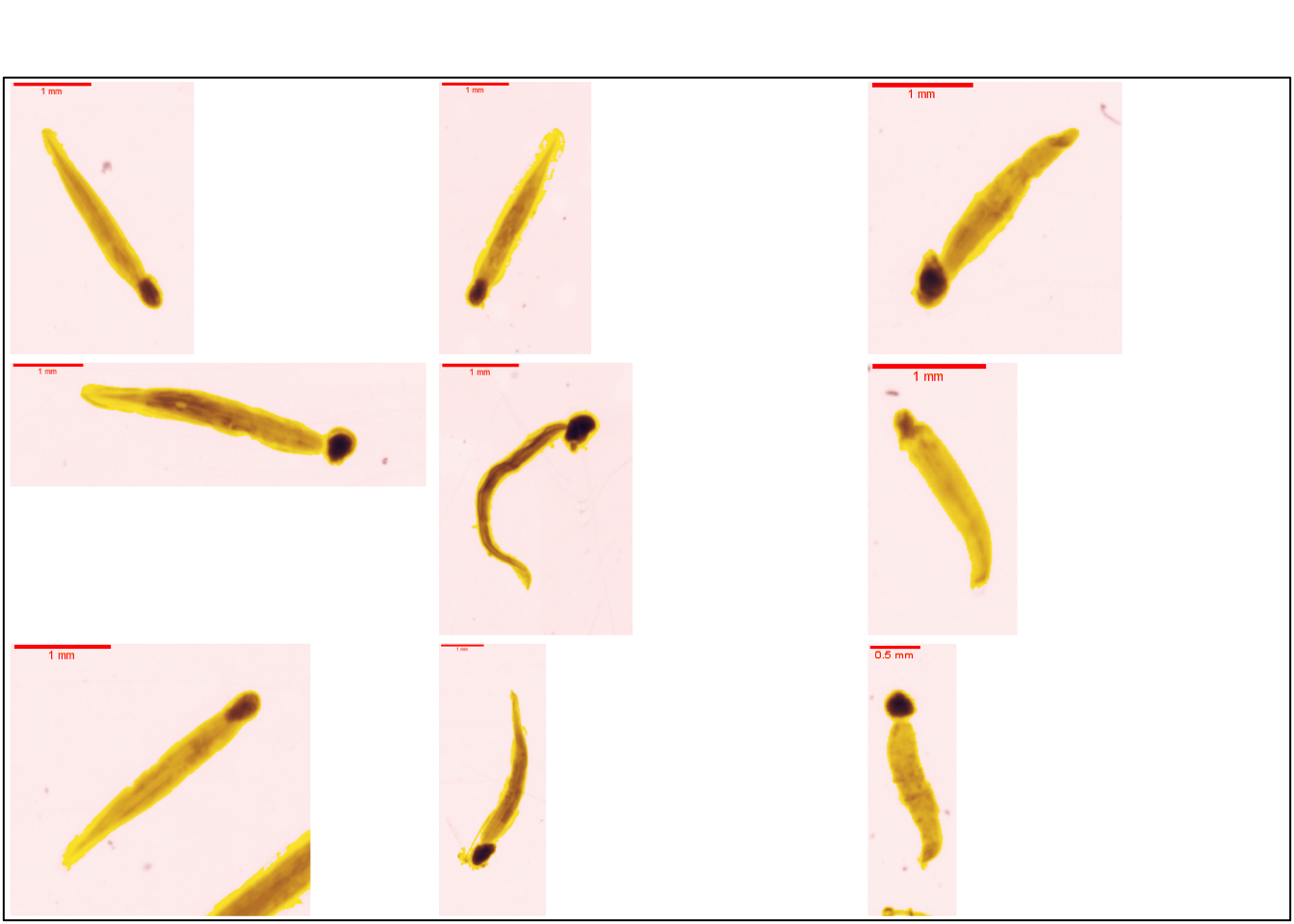
7.1.2.1.2 Fritillariidae
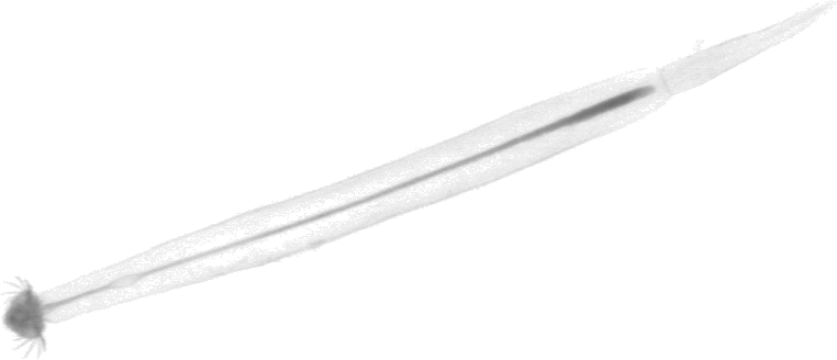
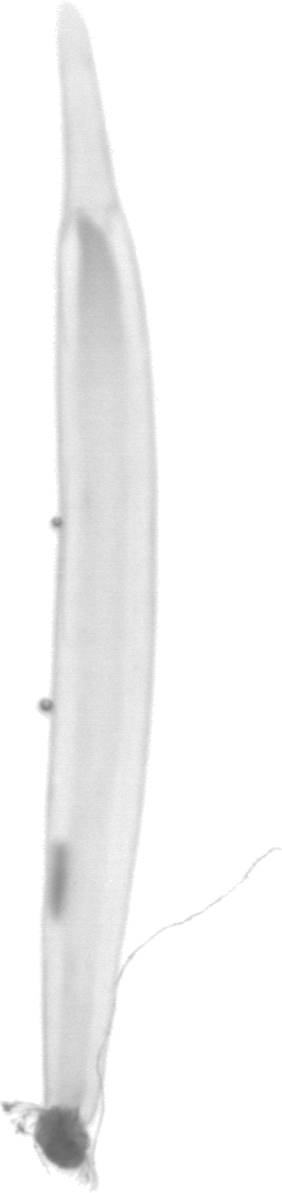
The trunk of these organisims is elongated. The trunk is less dense and compact than that of the OIkoploridae ( see Section 7.1.2.1.1 ). The colour of the tail of these organisms is rather light on the vignettes (Figure 7.11).
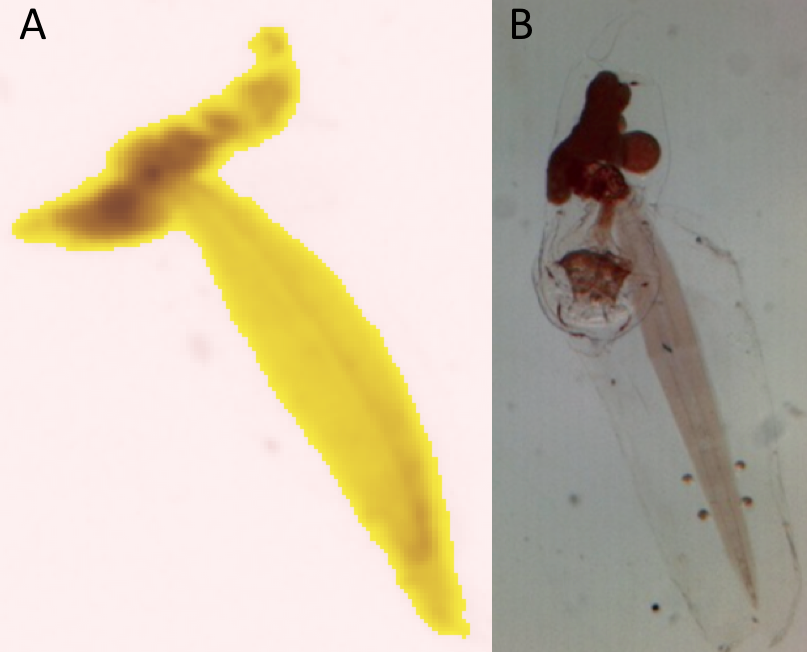
Figure 7.12 shows several vignettes of Fritillariidae.
plot_vignettes(vigns, group = "Fritillariidae")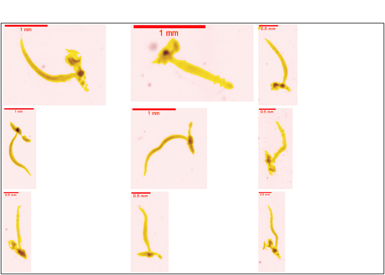
7.1.2.2 Appendicularia_tail
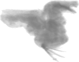
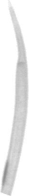
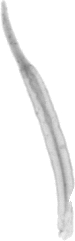
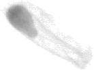
The organism is often damaged during the haul. As a result, appendicular tails are found in the samples (Figure 7.13). There is a light band arround two dark bands (Gorsky and Castellani 2017).
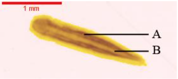
Figure 7.14 shows several vignettes of the Appendicularia tail.
plot_vignettes(vigns, group = "Appendicularia_tail")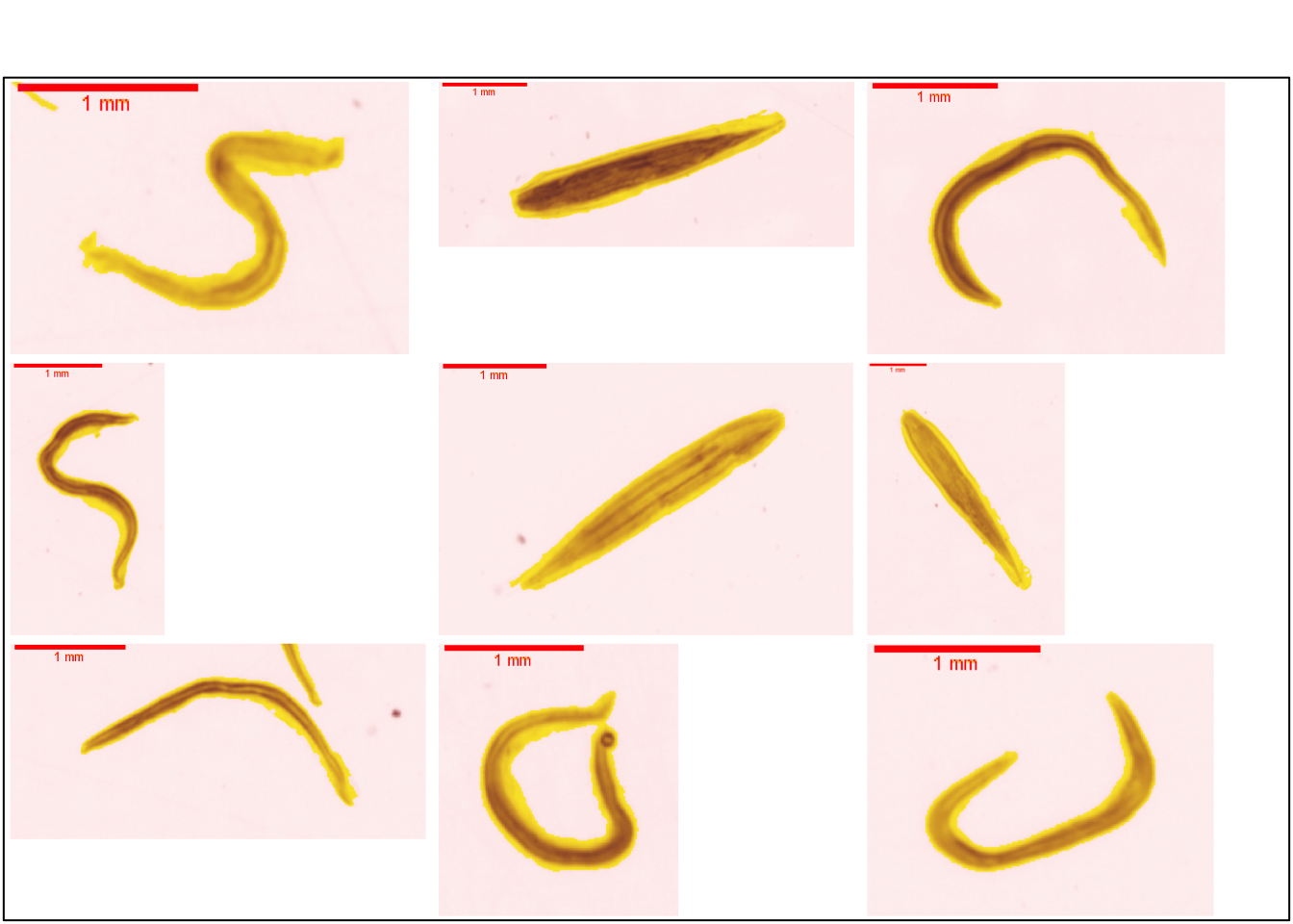
This group is similar to the tail__appendicularia group in ZooScanNet ( Figure 7.15 ).
7.2 Chaetognatha
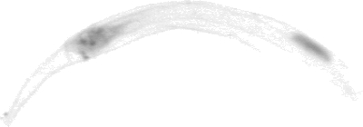
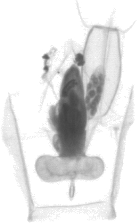
Chaetognatha are well studied pelagic predators. They are the main predators of copepods. Figure 7.16 shows the 3 characteristic parts of these organisms: head, trunk, tail (Pierrots-Bults 2017).
Fins are used to determine the species of chaetognaths (Pierrot-Bults 2020). The image analysis used to obtain the thumbnails does not allow sufficient accuracy of the fins to be maintained (Dugauquier 2019).
7.2.1 Sagittoidea
Phragmophora are distinguished from Aphragmophora by the presence of transverse musculature and abundant glandular structures on the body surface. Dugauquier (2019) further classified into Krohnittidae, Sagittidae and Eukrohniidae (see (Dugauquier, Engels, and Grosjean 2019) ). This discrimination is complex on the basis of vignettes.
Figure 7.17 shows several vignettes of Sagittoidea.
plot_vignettes(vigns, group = "Sagittoidae")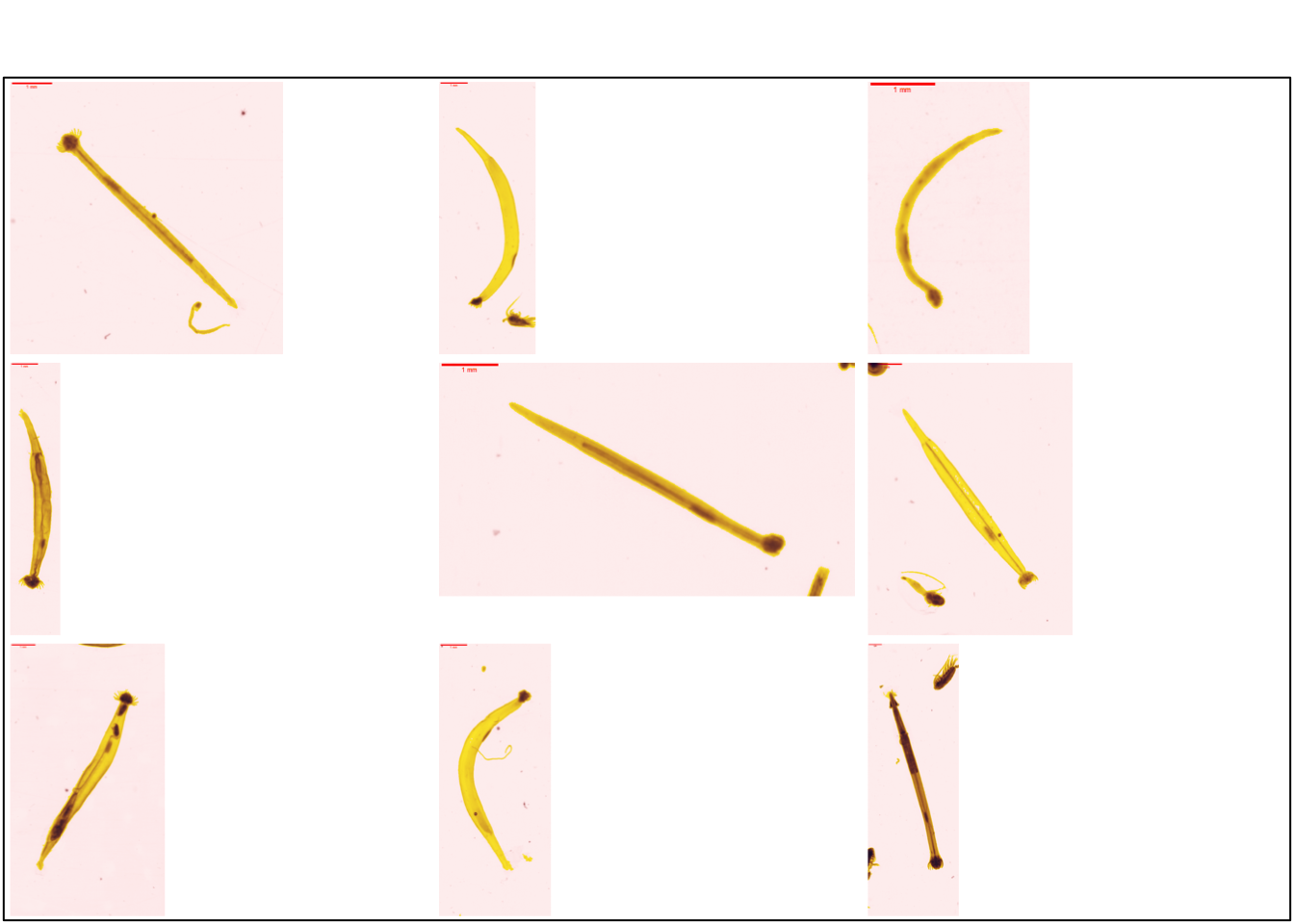
This group is similar to chaetognatha in ZooScanNet ( Figure 7.18 ).
7.2.2 Chaetognatha_head
New group
In previous studies (and associated training sets), this group was not studied.
During sampling, these organisms can be divided into fragments. The head is separated from the trunk and the tail ( Figure 7.19 ). This group includes the heads of the organisms named `head` in the ZooScanNet (Elineau et al. 2018).
7.2.3 Chaetognatha_tail
New group
In previous studies (and associated training sets), this group was not studied.
During sampling, these organisms can be divided into fragments. The head is separated from the trunk and the tail ( Figure 7.20)
In ZooScanNet the group that includes chaetognatha tails is called tail__chaetognatha (Elineau et al. 2018).

7.3 Ctenophora
New group
In previous studies (and associated training sets), this group was not studied.
The anatomical particularity of this group is the presence of 8 combs (bands composed of cilia) which allow the locomotion of the organism. These longitudinal bands are symmetrical all around the organism. This group is a rare group with a very limited number of images 39 in the ZooScanNet (Elineau et al. 2018).
7.4 Cnidaria
7.4.1 Scyphozoa
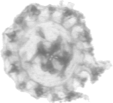
Schyphozoans are commonly known as jellyfish. These organisms are both benthic and pelagic (Licandro, Fischer, and Lindsay 2017). The successive stages of jellyfish development are presented below.
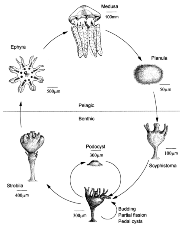
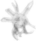
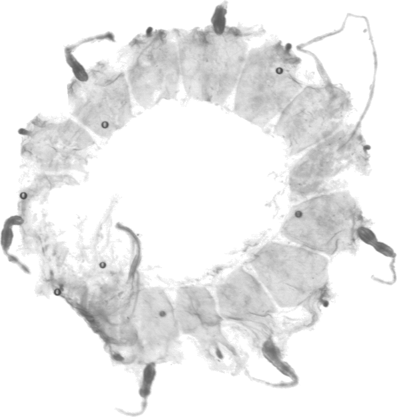
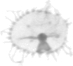
7.4.1.1 Scyphozoa_ephyra
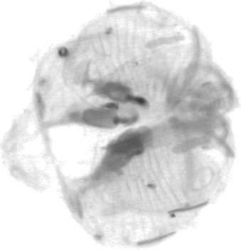
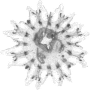
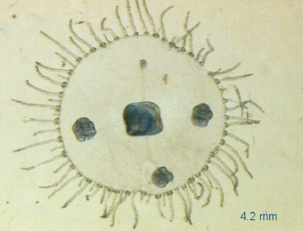
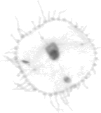
The ephyra stage is easily recognized by its 8 symmetry ( Figure 7.21 ). The radial and adriatic canals are darker in the vignettes. The morphology of the radial and adriatic canals can be used to refine the classification. However, we will not go any further in this book.
plot_vignettes(vigns, group = "Scyphozoa_ephyra")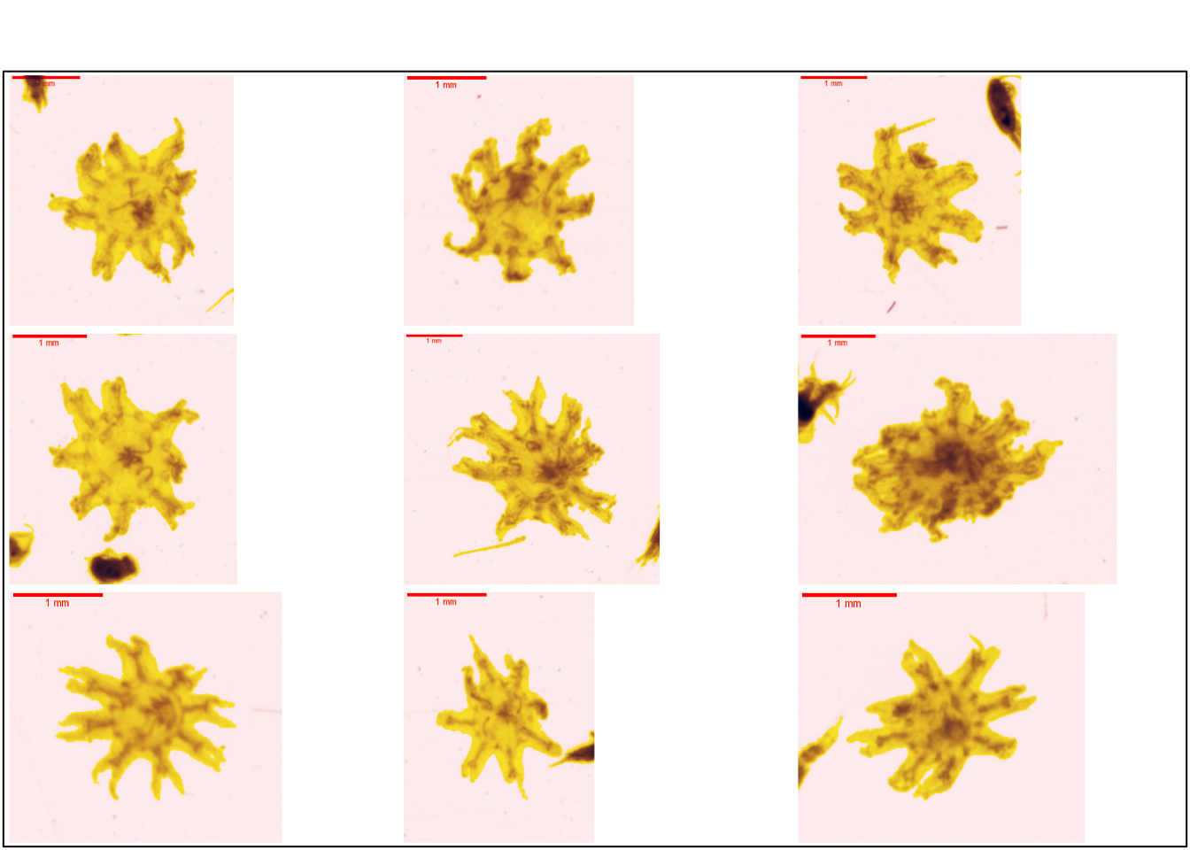
This group is similar to the ephyra group in ZooScanNet (Figure 7.22).
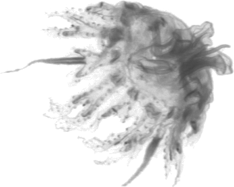
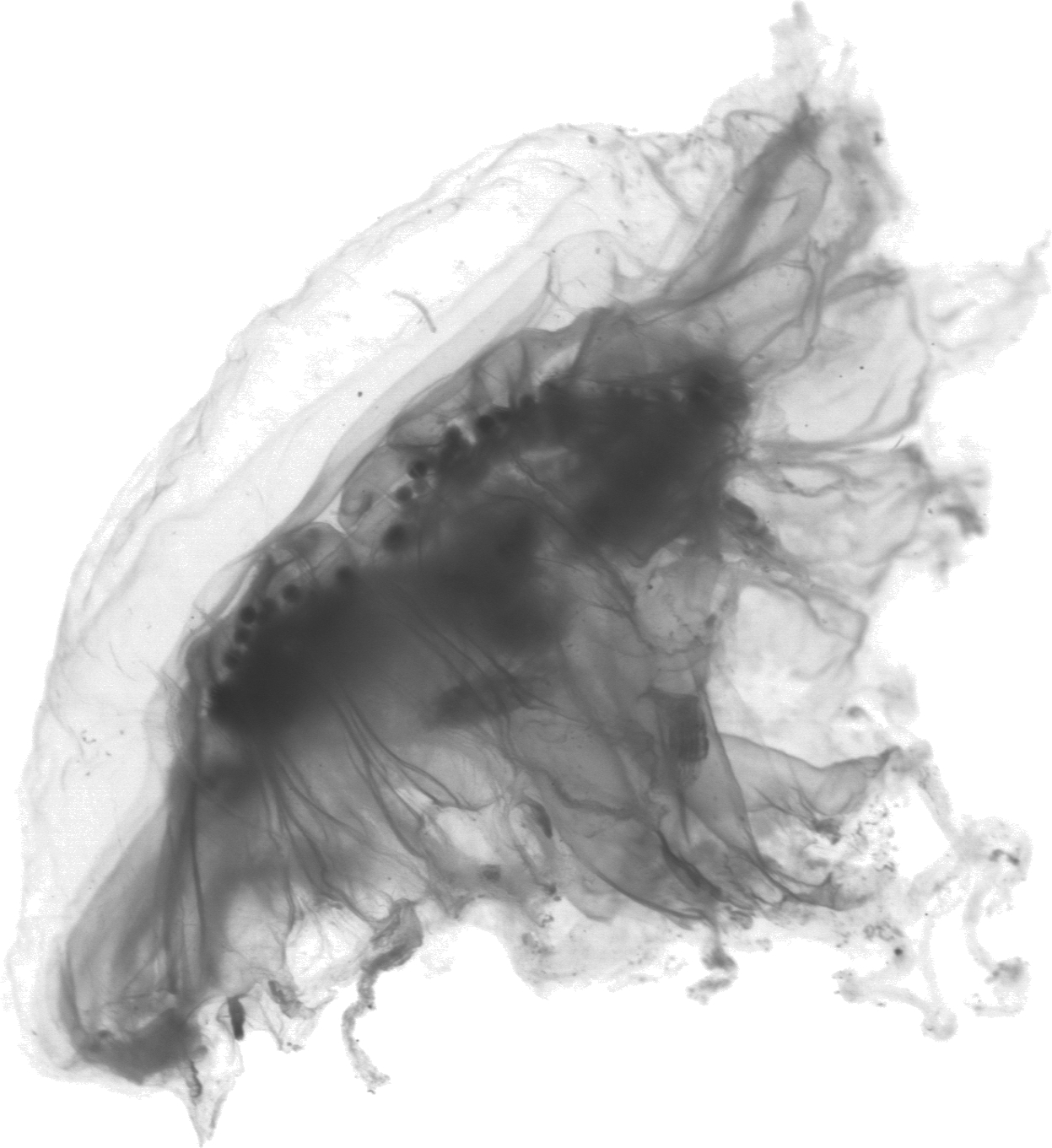
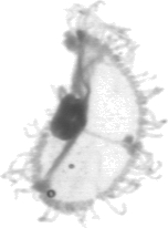
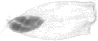
7.4.1.2 Scyphozoa_medusa
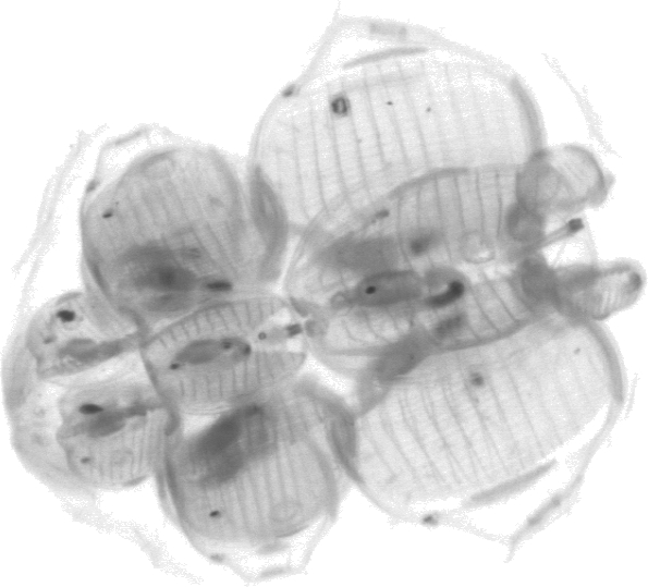
In the medusa stage, the symmetry of 8 is less marked. The individual canals are always larger than in the ephyra stage. The organisms are generally larger than those of the ephyra stage (Figure 7.23).

Figure 7.23 shows several vignettes of Scyphozoa.
plot_vignettes(vigns, group = "Scyphozoa_medusa", nx =2, ny = 2)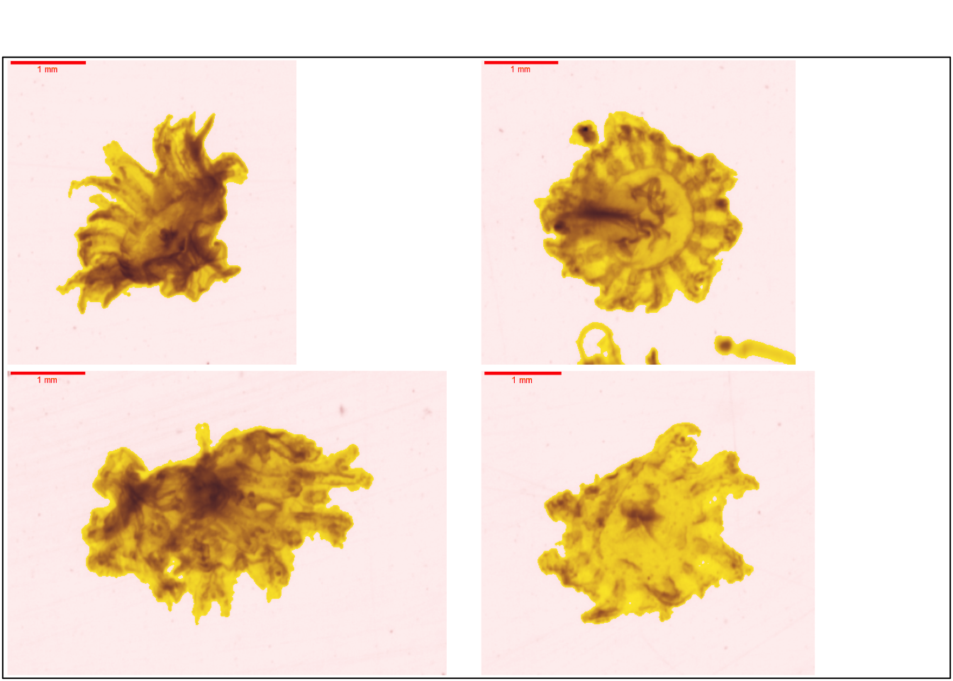
This group is similar to the scyphozoa group in ZooScanNet ( Figure 7.25 ).
7.4.2 Hydrozoa
7.4.2.1 Leptothecata
7.4.2.1.1 Campanulariidae
7.4.2.1.1.1 Obelia
New group
In previous studies (and associated training sets), this group was not studied.
In the centre, the mouth of the organism is connected by radial canals to the gonads. The circular canal and the tentacular bulbs are marked at the base of the tentacles ( Figure 7.26 ) .
This group is similar to the obelia group in ZooScanNet ( Figure 7.27 ).

7.4.2.2 Siphonophorae
Les siphonophores sont une colonie d’individus que l’on nomme zoïdes. On discrimine différent zoïdes par rapport à leur fonctions nectophore (locomotion), gastrozoïde ( digestion), pneumatophore (flottaison) (Licandro, Carré, and Lindsay 2017).
7.4.2.2.1 Abilidae
Les abylidae ne possèdent pas de nectophores tout comme les Diphydae.
7.4.2.2.1.1 Abylopsis_tetragona_eudoxie
La Figure 7.28 présente sur la partie supérieur ce que nous classons dans la catégorie Abylopsis_tetragona_eudoxie et la partie inférieur Abylopsis_tetragona_gonophore .
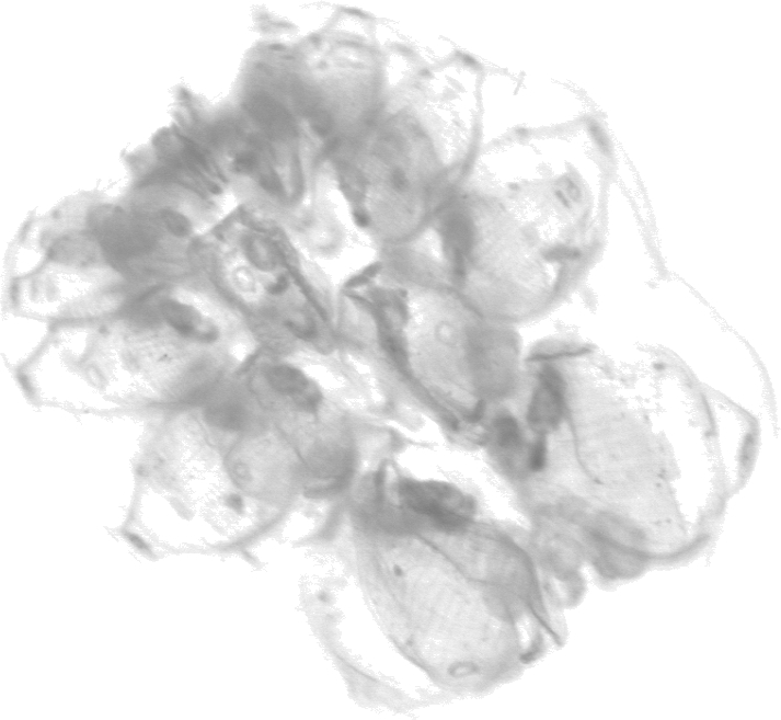
La Figure 7.29 présente plusieurs vignettes du stade eudoxie d’Abylopsis tetragona (partie bractée). La forme pentagonale est très marquée ainsi que les gastrozoides plus denses sur les vignettes.
plot_vignettes(vigns, group = "Abylopsis_tetragona_eudoxie")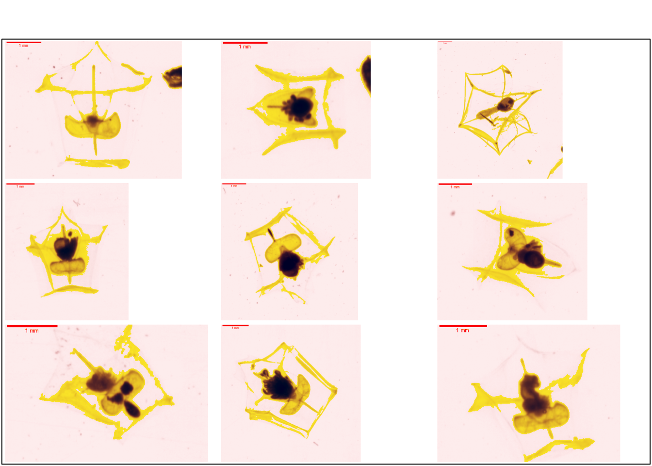
Ce groupe est similaire au groupe eudoxie__Abylopsis_tetragona dans le ZooScanNet ( Figure 7.30 ). Les deux premières images comprennent uniquement la partie bractée de l’organisme alors que les deux suivantes proposent la partie bractée et le gonophore.
7.4.2.2.1.2 Abylopsis_tetragona_gonophore
La partie inférieur de la Figure 7.28 présente les gonophores d’une forme de prisme rectangulaire. La Figure 7.31 présente plusieurs vignettes du gonophore d’abylopsis tetragona.
plot_vignettes(vigns, group = "Abylopsis_tetragona_gonophore", nx = 3, ny = 2)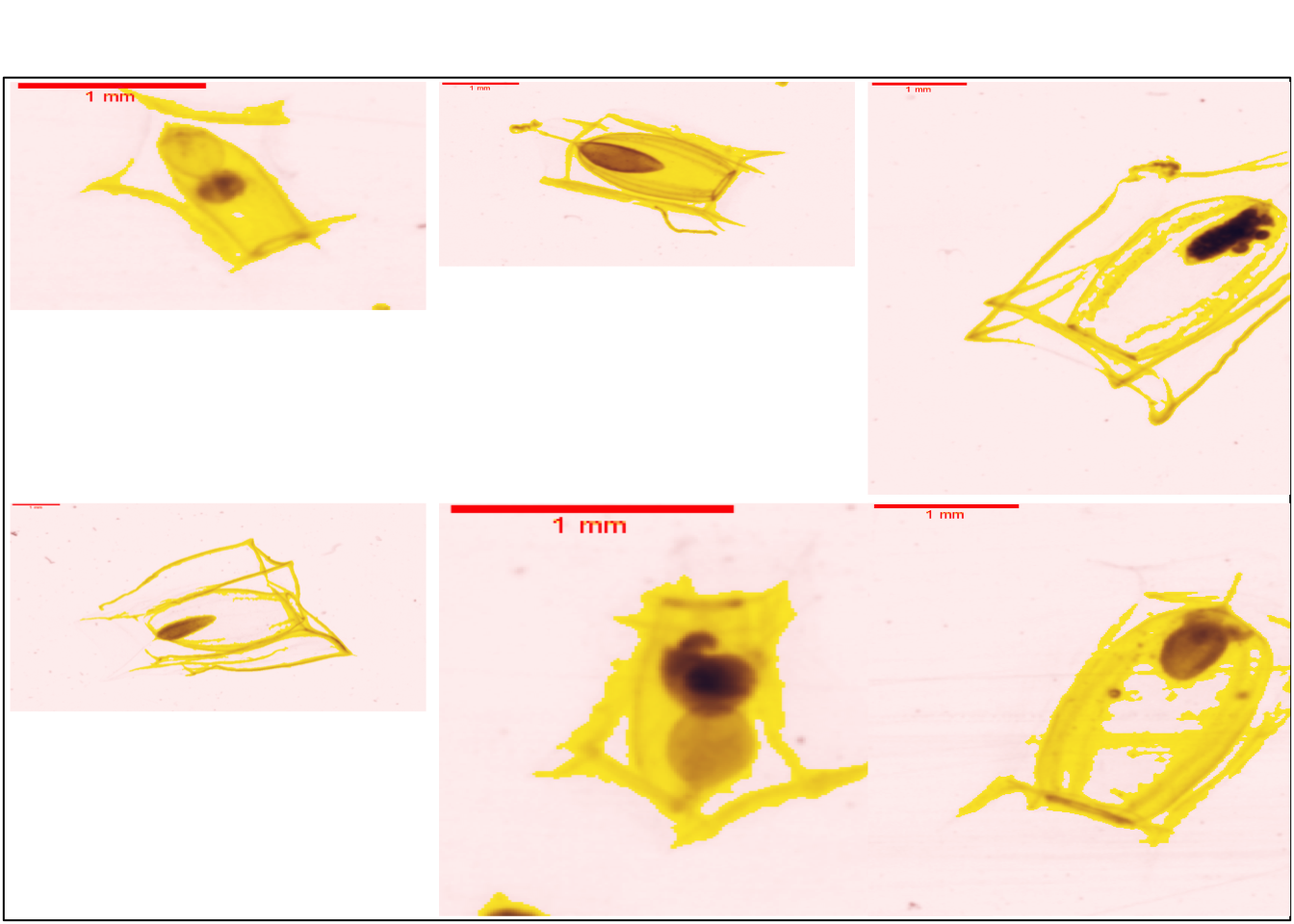
Ce groupe est similaire au groupe gonophore__Abylopsis_tetragona dans le ZooScanNet ( Figure 7.35 ).
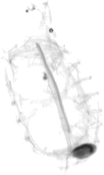
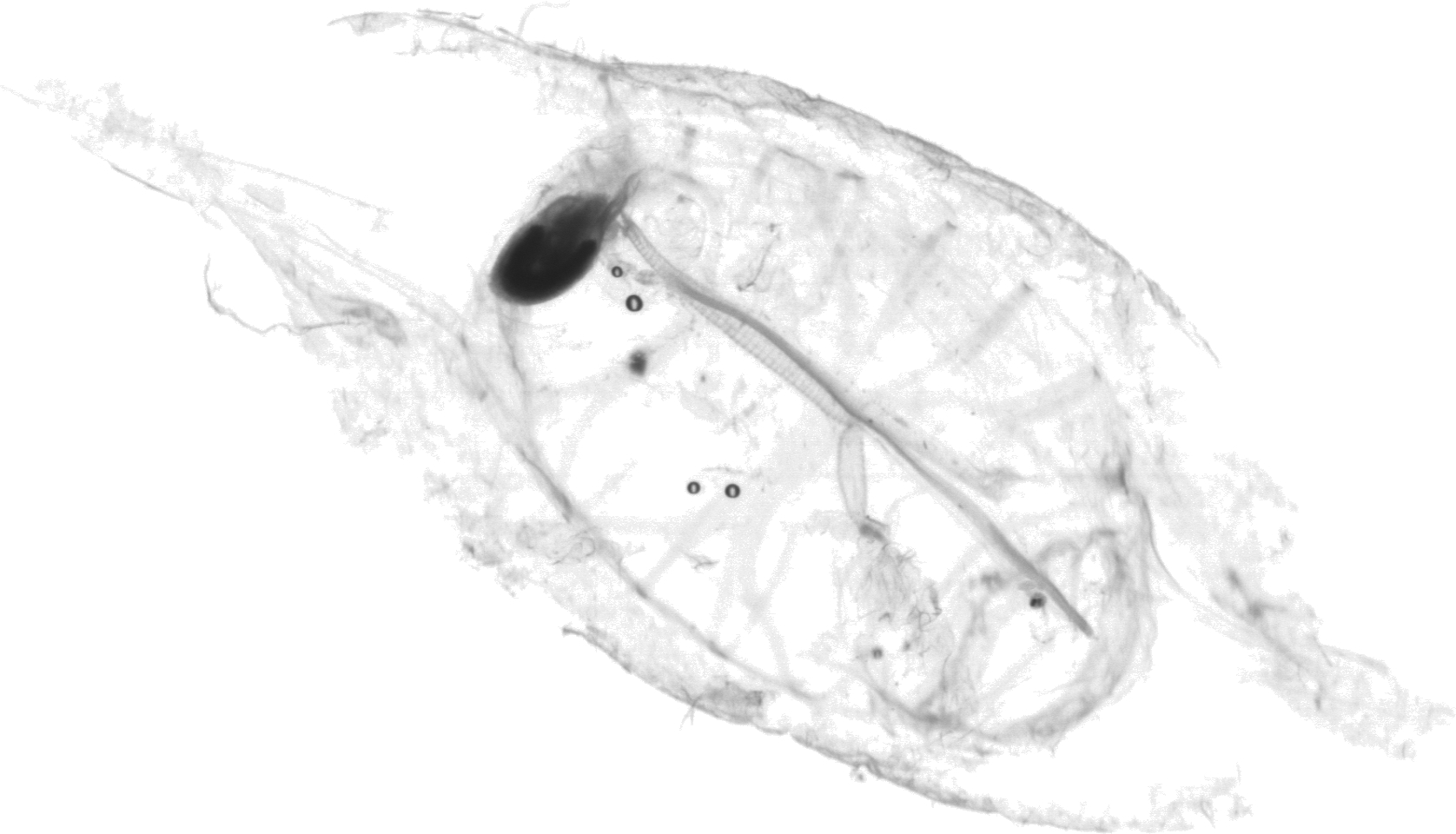
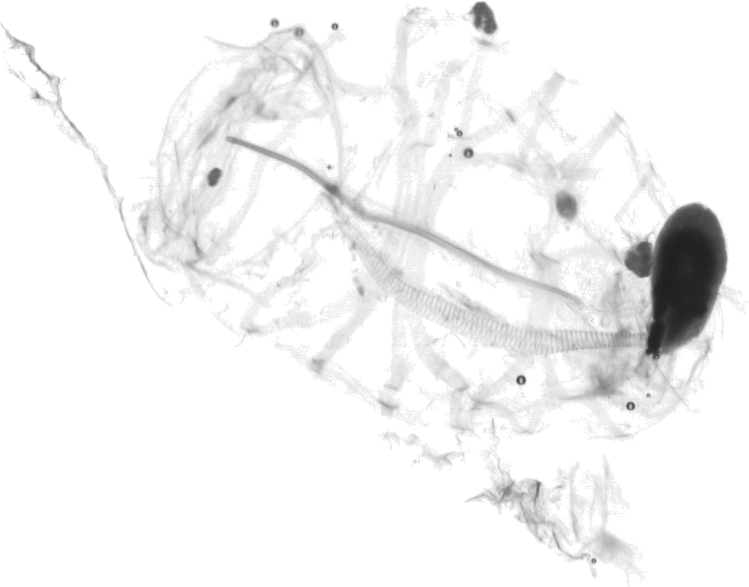
7.4.2.2.1.3 Abylopsis_tetragona_nectophore
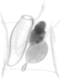
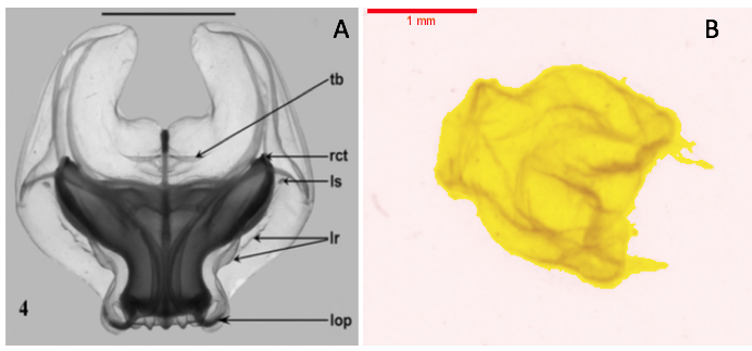
Plusieurs éléments sont identifiables sur le nectophore antérieur des Abylidée (Figure 7.33) :
la forme pentagonales
la cavité sous ombrellaire (plus grande des 3 cavités, claire sur les vignettes)
gastrozoide (cavité la plus dense sur l’image)
somatocyste (cavité ovale avec un petit diverticule)
La Figure 7.34 présente plusieurs vignettes du nectophore d’abylopsis tetragona.
plot_vignettes(vigns, group = "Abylopsis_tetragona_nectophore")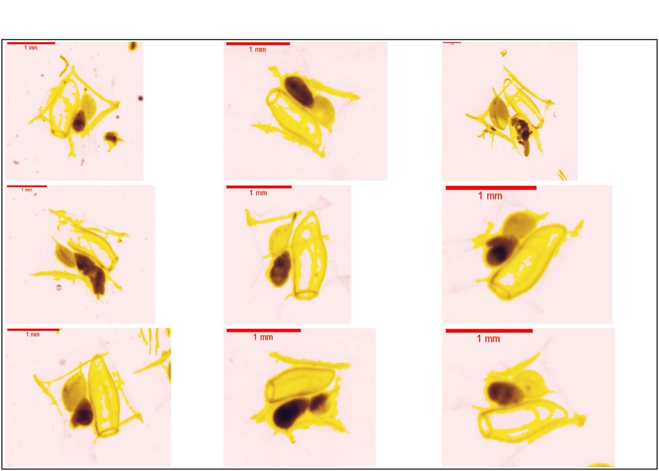
Ce groupe est similaire au groupe nectophore__Abylopsis_tetragona dans le ZooScanNet ( Figure 7.35 ).
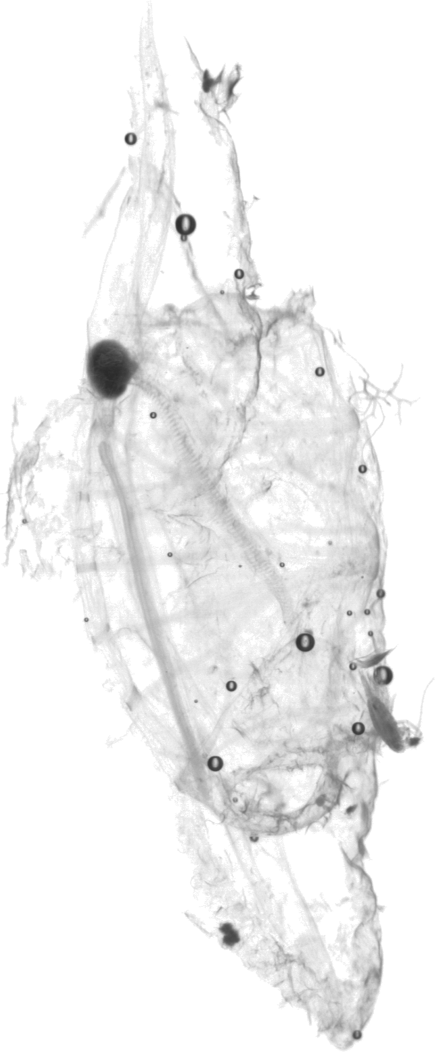
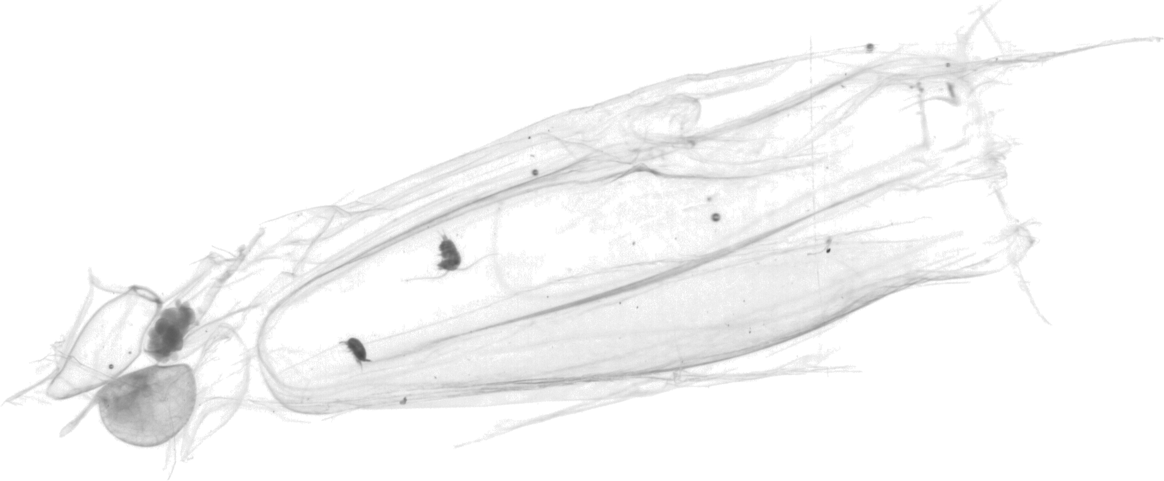
La Figure 7.36 propose une organisme complet doté de son nectophore antérieur et de son nectophore postérieur
7.4.2.2.2 Diphydae
7.4.2.2.2.1 Diphyidae_gonophore
La série de vignettes présente les gonophore qui sont la partie reproductrice du stade eudoxie. On retrouve une certaine ressemblance avec les gonophores d’Abylopsis tetragona sans le prisme rectangulaire ( Figure 7.37 ).
plot_vignettes(vigns, group = "Diphyidae_gonophore")
Ce groupe est similaire au groupe gonophore__Diphyidae dans le ZooScanNet ( Figure 7.38 ).
7.4.2.2.2.2 Diphyidae_nectophore
Les nectophores des Diphyidae sont de formes coniques. Le nectosac est plus densus sur les vignettes ci-dessous. Le long du nectosac, on observe les somatocystes ( Figure 7.39 ).
plot_vignettes(vigns, group = "Diphyidae_nectophore")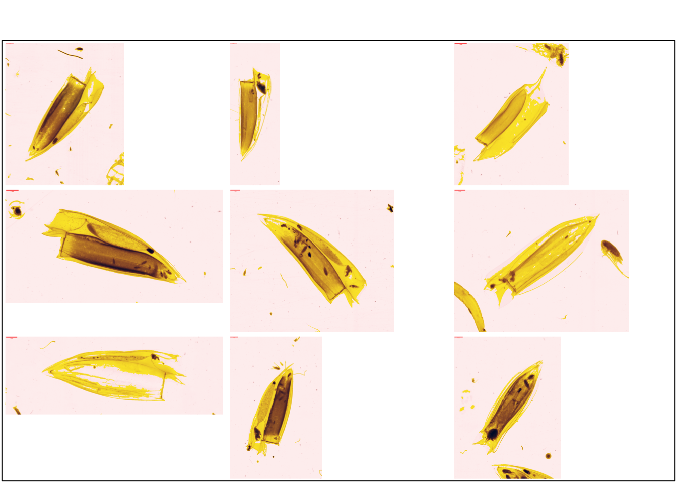
Note
Le ZooScanNet propose deux groupes : eudoxie__diphyidae et nectophore__diphyidae . Nous faisons le choix de les regrouper en un seul et unique groupe : Diphyidae_nectophore .
7.4.2.2.3 Physonectae
7.4.2.2.3.1 Physonectae_nectophore
Ces organismes sont constitués d’un pneumatophore contrairement aux Calycophore. La forme particulière de ces organismes les rendent aisément identifiables. On observe un resserrement à la base de la colonie.
La Figure 7.40 présente plusieurs vignettes du stade méduse.
plot_vignettes(vigns, group = "Physonectae_nectophore")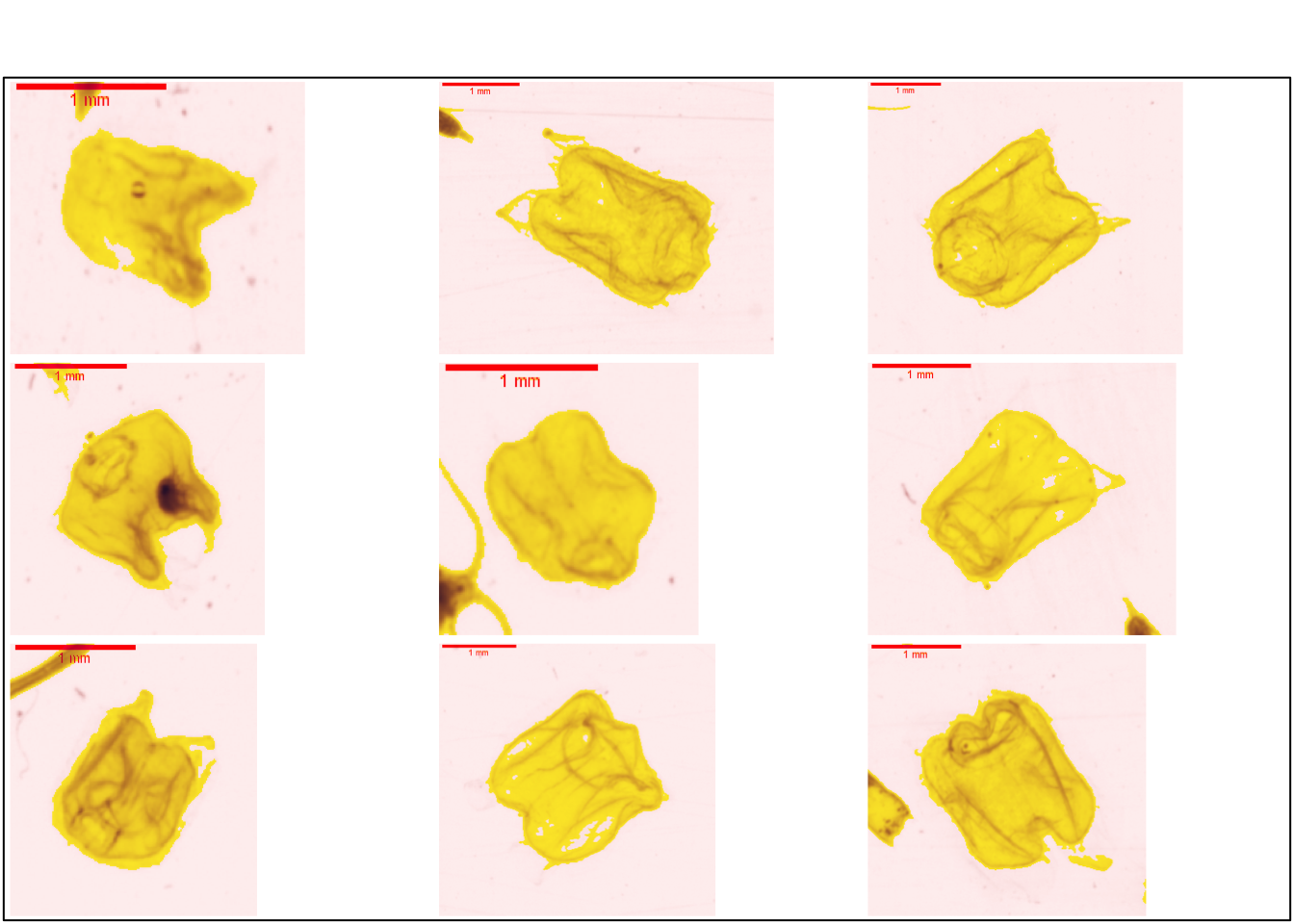
Ce groupe est similaire au groupe nectophore_Physonectae dans le ZooScanNet ( Figure 7.41 ).
7.4.2.3 Trachymedusea
7.4.2.3.1 Rhopalonematidae
7.4.2.3.1.1 Aglaura
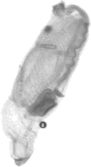
La Figure 7.42 présente plusieurs vignettes de Rhopalonematidae. La forme assez particulière de ce genre permet de l’identifier assez aisément. La partie plus dense dans la partie supérieur de l’organisme est son estomac et les ses gonages suspendue par un pédoncule.
plot_vignettes(vigns, group = "Aglaura")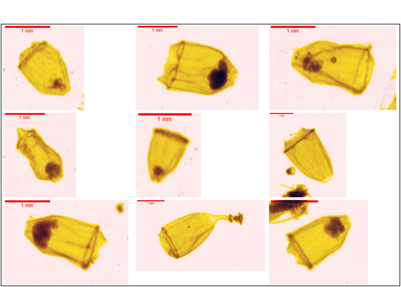
Ce groupe est similaire au groupe Aglaura dans le ZooScanNet ( Figure 7.43 ).